
Modern X-ray diagnostics

Wise people see everything.
Pola Negri
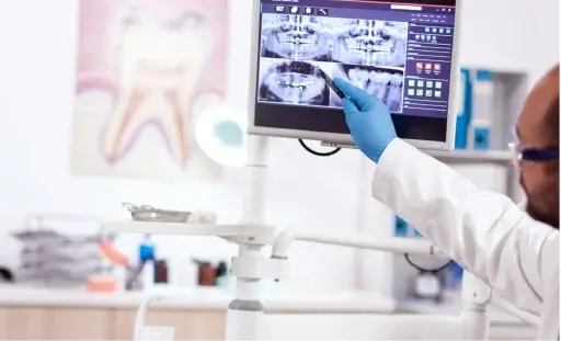
The deeper inside the forest, the darker it gets….this definitely doesn’t describe us. We can see everything because radiological examinations play an important part in dental diagnostics. This serious sounding name simply means an X-ray, yet a new dimension of an ordinary X-ray.
In our Clinic roentgen films went out of date. Now we’re closer to perfection. We use state-of-the-art digital programmes for taking dental radiographs by means of digital radiography techniques (the image of the examined object is obtained not on a roentgen film but on a computer screen), which decrease the radiation dose up to 9 times in comparison with traditional X-rays.
This is not the end of our novelties and innovations. Digital software connected to the dental panoramic radiograph and other radiographic devices allows for the display of more details on the screen, as well as for the image to be viewed in 3D. Thanks to X-ray diagnostics nothing will escape the dentist’s attention.
Success in dental treatment is ensured by the experience and knowledge of the dentist supported by the possibilities provided by modern diagnostic tools.
Our X-ray lab offers:
- panoramic x-rays,
- dental X-rays,
- sinus X-rays,
- temporomandibular joint X-rays and
- teleroentgenographic images required for orthodontic treatment.
And this means that nothing human is…invisible to us.
Our system of X-ray diagnostics includes not only an X-ray laboratory, but also radiographic devices that make it possible to produce images in surgeries without leaving the dentist’s chair.
Modern IT solutions make it possible to examine previously taken images directly in front of the patient and during treatment. They also enable the images to be processed for diagnostic and documentation purposes.
RVG (radiovisiography, point X-ray images of teeth, i.e. point X-rays) – digital radiography with minimal radiation dose, which can be used multiple times without risk to health in all patients, including children. Radiographic teeth images at Villa Nova are characterised by perfect quality.
This type of X-ray is applied in nearly all areas of dentistry: from detecting areas of caries in conservative dentistry to the assessment of the degree of implant osteointegration. Tooth X-rays provide the most information about the condition of single teeth and are characterised by the greatest accuracy in reproducing details.
An orthopantomogram (dental panoramic radiograph, so-called panorex) enables to show the entire dentition, jaw bones as well as neighbouring structures in one image, with a dose of radiation corresponding to 4-5 tooth x-rays. Despite the fact that an orthopantomogram is less accurate that single tooth X-rays, it allows for a comprehensive assessment of the status of dentition, which explains its significant usefulness in the diagnostic process, treatment planning and the assessment of results.
The projection geometry of panoramic X-rays ensures the highest quality, regardless of the anatomy of the facial skeleton. Other advantages of orthopantomograms include: no nuisance to the patient, the possibility of carrying out the examination in patients unable to open their mount, image understandable to the patient.
An orthopantomogram is essential to provide an accurate diagnosis. Based on the image, we can hold a consultation and present a treatment plan already during the first appointment, by assessing e.g. the condition of teeth, bones, the presence of inflammatory lesions, impacted teeth (e.g. wisdom teeth), sinus or periodontal diseases.
An orthopantomogram is essential inimplantological and orthodontic treatment. At Villa Nova Dental Clinic panoramic X-rays are taken routinely as a preliminary examination before the commencement of dental treatment.
Acephalometric radiograph (cephalogram or cephalometric X-ray) is a radiograph of the head, used particularly for orthodontic diagnosis. A detailed analysis of cephalograms allows for an assessment of spatial relationships of teeth, jaws and other skull structures in the patient. It is necessary for accurate diagnosis of occlusal abnormalities and treatment planning. The comparison of examinations performed before and after the treatment makes it possible to evaluate the effects of treatment.
Temporomandibular joint X-ray are an invaluable help in diagnosing conditions affecting these body structures. They constitute addition examinations in dental surgery, prosthetics, as well as orthodontics, because all these areas deal with the treatment of temporomandibular joints, though from different perspectives. Joint disorder symptoms are often subtle and ambiguous, while the radiological examinations may provide a lot of information about the cause, location and nature of the abnormality.
1. Are there any contraindications for a radiological examination?
The primary contraindication is pregnancy. A radiological examination during pregnancy should only be performed when health benefits from the examination clearly outweigh the risk of exposing the foetus to radiation. In case of an examination being performed during pregnancy, special shields protecting the foetus from radiation are used.
2. How often can digital radiographs (RVG) be taken?
Digital radiographs, similarly to other radiological examinations, should be limited to necessary cases, yet at the same time we need to be aware of the fact that modern equipment provides high quality of the examination with minimal radiation doses. Also, because it’s a digital technology, it practically eliminates the risk of a failed radiograph.
3. Is radiological diagnostics safe for children?
A diagnostic radiograph does not pose a direct threat to the child’s health, but the frequency of examination is an important factor. A minimal number of radiological examinations and the application of the latest methods for radiation dose reduction are recommended. Modern radiological devices offer special software that enables to significantly reduce the radiation and keep high image quality at the same time. Taking into account the safety of children at our Clinic, it is recommended to use digital radiography as a routine diagnostic examination, as this device is characterised by minimal harmful radiation.
4. When should a panoramic X-ray (“panorex”), and when a cephalometric X-ray be taken?
A panoramic X-ray is an image that provides a general overview of the teeth, the mandible, the maxilla and some part of the patient’s sinuses. Such X-ray should be taken at the beginning of the treatment, especially when the patient hasn’t visited a dentist for quite a while. A cephalometric X-ray is a specialist image used in diagnostics and establishing plan of the treatment of occlusal abnormalities (see -> orthodontics).
5. Why is it sometimes necessary to have additional digital radiographs/point X-rays taken despite having a recent panoramic X-ray (no older than 6 months)?
It happens because a panoramic X-ray is a general image, which provides an overview of the patient’s dentition. Point X-rays represent a significantly smaller area – usually several teeth, but in greater detail. Root canal treatment in particular requires a point X-ray before and after treatment (sometimes in the course of treatment as well). The radiographs are often taken from different angles, which allows the doctor to see all root canals in a tooth and confirm that the fillings are appropriate.
6. Can a radiological examination be performed in patients with pacemakers?
There are no contraindications for radiological examinations in patients with pacemakers. However, it needs to be borne in mind that image artefacts (distortions) can occur. This is because the metal, from which the pacemaker is made, absorbs roentgen radiation. It is not allowed to perform an MR scan in such patients, because the magnetic field can damage or destroy the pacemaker, as well as generate heat that can burn the patient.